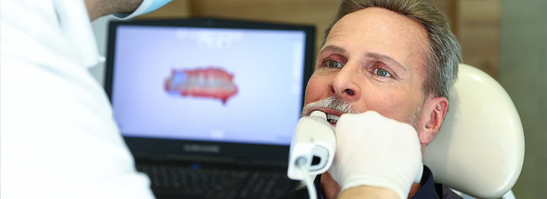
Digital impressions are high-resolution, computer-generated records of your teeth and surrounding oral tissues created with an intraoral optical scanner. Instead of traditional putty and trays, a handheld wand captures a rapid sequence of images or video that specialized software stitches together into a precise three-dimensional model. That model can be viewed immediately on-screen, rotated, and inspected from multiple angles to ensure the dentist has captured the necessary detail.
Different systems use slightly different scanning technologies—some capture hundreds of images per second while others use structured light or laser-based scanning—but the end result is the same: an accurate digital replica of the mouth that can be used for diagnosis, treatment planning, and fabrication of restorations. Because the model exists as a digital file, it can be archived, compared to prior scans, or transmitted electronically to a dental laboratory or in-office milling unit.
This approach replaces several manual steps in the traditional workflow. Where conventional impressions require physical materials to set and then be poured into stone models, digital impressions remove those intermediate steps. The immediate, on-screen feedback also lets the clinician identify and correct any missed areas in real time, improving the quality of the capture and reducing the likelihood of retakes.
For patients, the most noticeable difference is comfort. Many people find conventional impression materials unpleasant—some experience gagging, discomfort from rigid trays, or anxiety about having sticky substances in their mouths. A digital scan is performed with a small, smooth wand that glides over the teeth and gums, which most patients find far more tolerable.
Speed is another patient-centered advantage. Scanning sessions are typically shorter than traditional impression appointments, and because the dentist can immediately review the scan, there is less chance of needing a repeat visit for an inaccurate impression. For treatments that require a series of records—like orthodontic monitoring or progressive restorative work—digital files simplify follow-up visits and make tracking changes over time more convenient.
Digital impressions also reduce logistical delays. When a lab is involved, digital files are transmitted electronically rather than relying on physical shipments. That can speed up the process for restorations and shorten the overall treatment timeline. For patients who value efficiency and minimal interruption to their daily lives, these technology-driven improvements are especially beneficial.
One of the strongest clinical arguments for digital impressions is precision. Modern intraoral scanners capture fine surface detail and spatial relationships between teeth with a level of repeatability that supports well-fitting crowns, bridges, implant restorations, and veneers. A more accurate impression translates into restorations that seat more predictably, require fewer adjustments, and achieve better marginal adaptation.
Fewer adjustments at the chairside reduce appointment time and can improve long-term results by minimizing repeated removals and reseating of provisional restorations. For implant-supported work, the ability to merge digital impressions with cone-beam CT data enables more precise planning and prosthetic integration, which helps align the restorative and surgical phases of care for a more predictable outcome.
Digital workflows also support contemporary prosthetic options such as same-day ceramic restorations produced in-office. When a digital impression is combined with CAD/CAM milling or 3D printing, it becomes possible to design and fabricate a definitive or temporary restoration within a single visit. This capability can preserve tooth structure and streamline treatment while maintaining high aesthetic and functional standards.
Finally, the digital record itself is valuable for monitoring and continuity of care. Scans can be archived and compared over time to track wear, movement, or changes in tissue contours, giving clinicians another objective tool to support preventive care and long-term treatment planning.
The digital workflow begins chairside with image capture. After an initial clinical assessment, the clinician uses the scanner to record the target area—this may be a single tooth preparation, a full-arch record, or an occlusal registration. The software processes the captured data instantly, creating a 3D model that can be refined, annotated, and used to generate a prescription for the laboratory or for in-office fabrication.
When a laboratory is involved, the digital file is exported in a standardized format and transmitted electronically. Technicians import that file into their CAD software to design the restoration, then manufacture it either by milling or additive 3D printing depending on the material and laboratory capabilities. For practices with in-office CAD/CAM equipment, the design can be completed in the same visit and sent directly to a milling machine to produce a ceramic crown or other restoration.
Integration with other technologies further enhances the workflow. Digital impressions can be merged with CBCT scans for implant planning or combined with orthodontic software for aligner fabrication. This interoperability reduces manual steps, improves communication between the clinician and lab, and supports a predictable, coordinated treatment plan.
The net effect is a streamlined clinical pathway: accurate data capture, efficient digital communication, and controlled fabrication that together reduce uncertainty, shorten turnaround time, and improve the likelihood of a first-time fit.
Digital impressions are a versatile tool, but there are practical considerations patients should understand. Some clinical situations—such as impressions that must capture deeply subgingival margins or a very large full-arch case—may present greater technical demands and require specific scanner techniques or adjunctive steps. Your clinician will determine whether a digital approach is appropriate for your particular procedure.
Preparation is minimal on the patient’s part. Keeping teeth clean and dry during the scan helps the software capture clearer data, so a routine cleaning before an extensive restorative appointment can be helpful. The clinician may use gentle retraction or a drying agent to expose margins and ensure an accurate capture of critical areas.
Because intraoral scanners and associated software are continually evolving, outcomes depend on a practiced clinical team and current equipment. Your dentist will choose the scanner and workflow that best fit the treatment objectives. If an in-office same-day restoration is planned, expect a coordinated sequence of digital design and milling steps; if a laboratory-fabricated restoration is preferred, the practice will transmit files and plan for a follow-up appointment for final seating.
Digital impressions represent a modern, patient-friendly advance in restorative and prosthetic dentistry. By replacing traditional materials with high-precision optical capture and digital workflows, this technology improves comfort, enhances accuracy, and streamlines communication between the dental team and the laboratory. At our Winter Park office, we use intraoral scanning as part of an evidence-based approach to deliver predictable restorative and cosmetic results. Contact Fay Hu General Dentistry to learn more or to discuss whether digital impressions are right for your treatment plan. Please contact us for more information.

Digital impressions are computer-generated, three-dimensional images of the teeth and surrounding oral tissues created with an intra-oral optical scanner. The scanner captures a sequence of high-resolution images that software combines into an accurate digital model. These digital models serve the same purpose as traditional putty impressions but are available instantly for review and verification.
The digital file can be viewed, measured and adjusted on-screen to confirm margins, contacts and occlusion before any laboratory or milling work begins. Because the model is electronic, it can be shared immediately with a dental laboratory or exported to in-office fabrication systems. This capability streamlines treatment planning for crowns, bridges, implant restorations and orthodontic devices.
Digital impression scanners use optical technology such as structured light, laser or confocal imaging to capture the shape of teeth and gums. The device records many small frames per second as the wand moves through the mouth, and specialized software stitches those frames into a continuous 3D model. Real-time feedback on the monitor helps the clinician identify areas that need additional scanning before the patient leaves the chair.
After capture, the software processes the data to refine surfaces, remove noise and generate a printable or exportable file format like STL. The finished file can be used for digital design, virtual articulation, or sent electronically to a lab for fabrication. This digital workflow reduces handling errors associated with physical impressions and improves communication across the care team.
Digital impressions improve patient comfort by eliminating heavy impression trays and dental putty, which can trigger gagging or discomfort. They also reduce the need for repeat impressions because the clinician can assess detail and completeness immediately on-screen. For the dental team, digital files simplify storage and retrieval compared with physical models and plaster casts.
From a laboratory and fabrication perspective, digital files transmit instantly and avoid shipping delays, handling damage or deformation that sometimes occurs with poured models. The digital workflow often results in more consistent fit and fewer adjustments at insertion. Overall, the process enhances precision, efficiency and predictability for many restorative and orthodontic procedures.
Digital impressions are generally more comfortable than traditional putty impressions because they use a small, handheld scanning wand rather than full-arch trays and material. The wand is maneuvered gently around the mouth and typically requires only short scanning passes to capture the necessary anatomy. Most patients report less gag reflex and reduced anxiety compared with conventional impression methods.
As for safety, intra-oral scanners are noninvasive and do not expose patients to ionizing radiation; they rely on visible or near-infrared light for image capture. The devices are cleaned and disinfected according to infection-control protocols between patients to maintain clinical safety. Clinicians also take care to avoid prolonged contact in sensitive areas and to pause scanning if a patient experiences any discomfort.
Modern digital scanners produce highly accurate impressions suitable for a wide range of restorations, including crowns, bridges and implant components. Accuracy depends on proper scanning technique, scanner calibration and software processing, but when used correctly the resulting digital models meet or exceed the precision of many traditional impression workflows. Clinicians can immediately evaluate margins, contacts and occlusal relationships to reduce the chance of remakes.
Laboratories that accept digital files are accustomed to working with STL or similar formats and can fabricate restorations with tight tolerances from these models. For complex or full-arch cases, clinicians may combine digital scans with additional records such as bite registrations or CBCT data to ensure the highest level of reliability. Continual software and hardware improvements further enhance reproducibility over time.
Yes. Digital impressions are a key component of same-day restorative workflows that use in-office design and milling systems to produce ceramic crowns, onlays and veneers during a single visit. After capturing the digital scan, the dentist or a trained technician designs the restoration using CAD software and sends the design to an in-office milling unit or 3D printer. This integrated approach reduces the need for temporary restorations and multiple appointments.
At Fay Hu General Dentistry the team uses digital imaging and in-office fabrication tools to streamline same-day care when clinically appropriate. Clinicians still evaluate case suitability based on tooth condition, occlusion and esthetic goals, and they follow established protocols to confirm fit and function before finalizing the restoration. When indicated, same-day restorations provide efficient, predictable outcomes with the convenience of fewer visits.
For implant therapy, digital impressions capture the position of implant scan bodies or surrounding dentition to create precise surgical guides and prosthetic components. The digital data can be merged with CBCT scans to plan implant placement in three dimensions and to design implant-retained crowns or hybrid prostheses. This digital integration improves communication between surgeon, restorative dentist and laboratory for coordinated implant workflows.
In orthodontics, digital models enable virtual treatment planning, simulation of tooth movements and fabrication of clear aligners or custom appliances. Scans provide accurate records of tooth position that can be used to track progress and make adjustments throughout treatment. The ability to share digital models quickly with labs or aligner manufacturers accelerates treatment initiation and helps maintain continuity of care.
During a digital impression appointment the clinician will prepare the teeth as required, isolate the area and then use the scanning wand to capture the arches or specific teeth. The process typically takes only a few minutes per arch, and the clinician will monitor the image on a screen to ensure complete coverage. If any areas require additional detail, the clinician can rescan them immediately to avoid remakes.
After scanning, you may be shown the digital model so the clinician can explain treatment steps or expected outcomes. The digital file is then processed and used for design, communication with the laboratory or in-office fabrication. Patients can expect a cleaner, quicker experience compared with traditional impression techniques.
Digital impression files are saved in standardized formats such as STL and can be stored securely on the practice's local server or in encrypted cloud systems used by dental laboratories and software vendors. Clinicians follow data security and privacy practices to protect patient information during storage and transmission. Access controls and secure transfer protocols help ensure that only authorized team members and laboratory partners can retrieve patient files.
When sharing files with a laboratory or specialist, the practice typically sends the digital data electronically, which eliminates shipping delays and reduces the risk of physical damage. Electronic transfer also creates an audit trail for case communication and can include annotated instructions for margins, materials and occlusal adjustments. These workflows improve coordination while maintaining record integrity.
While digital impressions have broad applicability, there are clinical situations where conventional impressions may still be preferred or used as an adjunct. Cases involving extensive soft-tissue management, certain full-arch prosthetic workflows, or laboratories with specific material or manufacturing requirements may benefit from traditional techniques. Additionally, clinicians sometimes capture a conventional impression when specific tissue-handling characteristics are necessary for a particular restoration.
Rather than replacing traditional methods entirely, digital and conventional workflows often complement one another based on case needs and practitioner judgment. The dental team will recommend the approach that best ensures an accurate fit, optimal function and long-term success for the patient. Patients should discuss any questions about technique choice with their clinician so they understand the rationale for the selected method.

We are dedicated to providing the highest quality of dental care to our patients.
Through excellence in dentistry and quality in relationships, we strive to positively impact your oral health, aesthetics, and self-esteem. From the front desk to the treatment room, our experienced team is here to support you with expert care and genuine compassion.