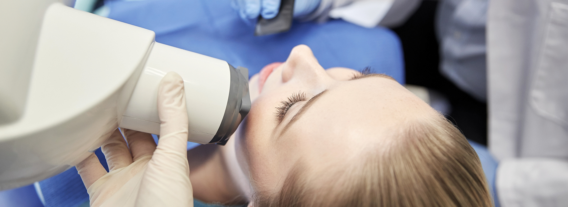
Digital radiography replaces traditional film with electronic sensors and computer processing to capture dental images. Instead of developing film with chemicals, the sensor records the X-ray and transfers it instantly to a computer where the image appears on a monitor. This shift from film to digital streamlines the imaging process and allows clinicians to evaluate teeth, bone, and supporting structures with greater speed and convenience.
For patients, the most noticeable change is the reduced waiting time: images are available immediately, so decisions about next steps can often be made during the same appointment. The digital file format also makes it easier to archive images in a patient’s chart, compare current and prior studies, and maintain a clear, searchable record of oral health over time. These efficiencies support both routine care and more complex treatment planning.
Digital radiography encompasses a range of technologies, from intraoral sensors used for bitewing and periapical images to extraoral panoramic units and cone beam CT systems for three-dimensional imaging. The core advantage remains the same — precise image capture combined with instant access and reliable storage — which improves the overall coordination of dental care.
One of the key advantages of digital sensors is image quality and flexibility. Digital images can be enhanced with software tools that adjust contrast, brightness, and magnification to reveal subtle details that might be missed on film. This level of control helps clinicians detect early-stage decay, evaluate root and bone anatomy, and identify changes in periodontal health with greater confidence.
Because digital files can be viewed at different levels of magnification without losing clarity, clinicians can examine small areas closely while still keeping sight of the broader anatomic context. That capability is particularly useful for endodontic assessments, implant planning, and evaluating fractures or resorption. Clearer visualization supports more accurate diagnoses and more predictable treatment planning.
In addition to on-screen enhancements, digital systems often include measurement tools that assist with implant positioning and restorative design. When combined with other digital workflows — such as intraoral scanning and CAD/CAM restorations — radiographic images become an important part of a coordinated, technology-driven approach to modern dentistry.
Digital radiography substantially lowers radiation exposure compared with conventional film X-rays because modern sensors are more sensitive and require less radiation to produce diagnosable images. Dose reduction is an important consideration for routine exams, pediatric patients, and those requiring periodic follow-up imaging. Clinicians follow the ALARA (As Low As Reasonably Achievable) principle while tailoring X-ray frequency and scope to each patient’s individual needs.
Beyond sensor efficiency, digital workflows allow targeted imaging strategies. For example, bitewing images that focus on detecting decay between teeth or periapical views that assess root tips can be captured selectively rather than taking wide-field exposures unnecessarily. When three-dimensional imaging is indicated, clinicians weigh diagnostic benefit against exposure and select appropriate field-of-view settings to limit radiation to the relevant area.
Because images are acquired and reviewed immediately, retakes are minimized. If an image needs to be repeated due to positioning or clarity, the adjustment can be made on the spot and the new image captured right away, further limiting exposure over time while ensuring diagnostic quality.
Digital radiography simplifies how images are stored, shared, and integrated into electronic health records. Once captured, images become part of the patient’s digital chart and can be archived, annotated, and retrieved without the physical limitations of film. This integration supports continuity of care by providing a clear, chronological view of dental findings and interventions.
Sharing images with specialists, labs, or referring providers is straightforward: a digital file can be transmitted securely for consultation or treatment planning. This capability speeds collaborative workflows for cases that require multidisciplinary input — for instance, coordinating between a restorative dentist and an oral surgeon when planning implant placement or complex extractions.
Digital records also support long-term monitoring. Clinicians can compare current images to prior studies side-by-side, making it easier to track progression or healing over months and years. This longitudinal perspective helps inform preventive strategies and timely interventions.
From a patient perspective, digital radiography enhances comfort and convenience. Modern sensors are designed to be thin and more comfortable than bulkier film holders, and shorter appointment times reduce chair time. Immediate image review also supports clearer communication: clinicians can show patients what they see on-screen, explain findings in real time, and include visual evidence as part of the discussion about oral health and treatment options.
Digital imaging is also kinder to the environment because it eliminates the need for chemical developers, fixers, and paper-based processing associated with film. Reducing chemical waste and physical storage needs is a practical advantage for any healthcare practice mindful of sustainability and efficient resource use.
Finally, the adaptability of digital systems makes them future-ready. As dentistry increasingly moves toward integrated digital workflows — combining scans, 3D imaging, and in-office milling or 3D printing — high-quality radiographic data becomes a foundational element for predictable, efficient care.
Our team incorporates digital radiography into routine exams and more advanced diagnostics to provide clear, timely information that supports clinical decision-making. Images captured during your visit are reviewed with you, explained in plain language, and stored securely within your chart so they can inform preventive care and any necessary treatment planning going forward.
Digital imaging is part of a broader technology portfolio used to enhance accuracy and patient comfort, from intraoral scanning to chairside restoration workflows and digital treatment planning. When higher-resolution or three-dimensional images are required, our clinicians select the modality that best balances diagnostic need and patient safety to ensure appropriate, personalized care.
By combining advanced imaging with careful clinical assessment, we aim to detect concerns early, plan treatments more precisely, and keep patients informed throughout the process. Fay Hu General Dentistry integrates these tools to deliver consistent, evidence-based care in a patient-centered environment.
In summary, digital radiography offers faster results, improved image quality, lower radiation exposure, and easier collaboration — all of which support better clinical decisions and a smoother patient experience. For more information about how digital imaging is used in our office and what to expect at your next visit, please contact us to learn more.

Digital radiography uses electronic sensors and computer processing to create high-resolution dental x-ray images that can be viewed, enhanced and stored digitally. Instead of traditional film, a small electronic sensor captures the image and transmits it immediately to a computer workstation. The resulting files can be enlarged, adjusted for contrast and examined in fine detail to help clinicians identify dental issues more accurately.
Because images are digital they integrate seamlessly with electronic health records and other dental technologies such as CBCT and digital impressions. This streamlines recordkeeping and allows clinicians to compare current images with prior studies to monitor changes over time. Many practices now rely on digital radiography as a standard imaging modality for both routine and complex dental care.
Traditional film x-rays require chemical processing and physical storage, while digital radiography produces instant images that are stored electronically. Digital sensors eliminate the need for film and development chemicals, which reduces environmental impact and simplifies workflow. The immediate availability of images also allows for faster clinical decision-making during the same appointment.
Digital images can be enhanced with software tools to clarify anatomy and reveal small changes that might be harder to see on film. Multiple clinicians can view the same image simultaneously, which supports collaboration and coordinated care. The ability to archive and retrieve images quickly reduces administrative burden and speeds follow-up evaluations.
Digital radiography typically requires significantly less radiation than conventional film radiography because modern sensors are more efficient at capturing x-ray photons. Dose-reduction protocols and updated equipment further minimize exposure while still producing diagnostically useful images. Clinicians also follow the principle of ALARA, keeping radiation doses as low as reasonably achievable for every patient.
Safety measures such as lead aprons and properly calibrated machines add layers of protection during imaging. Dentists evaluate each patient's needs and take x-rays only when clinically indicated, balancing diagnostic benefit with safety. For patients with concerns about exposure, the care team can explain the specific safeguards in place and the reason for the requested images.
Digital sensors are compact electronic devices placed inside or outside the mouth to capture x-ray photons and convert them into digital signals. The sensor's active element, often a charge-coupled device or complementary metal-oxide-semiconductor, records the pattern of x-ray attenuation produced by the teeth and surrounding tissues. That signal is then processed by software to generate a viewable image on a computer screen within seconds.
Sensors are designed to be durable and to provide consistent image quality across different exposure settings. They come in sizes and shapes suitable for pediatric and adult patients and can be positioned to capture periapical, bitewing or occlusal views. Proper sensor placement and patient cooperation are important to reduce retakes and ensure diagnostically useful images.
Digital radiography is used for a broad range of intraoral and extraoral imaging, including bitewing, periapical and panoramic studies. In many practices the same digital workflow integrates with cone beam computed tomography (CBCT) for three-dimensional planning, as well as with digital impressions and CAD/CAM systems. This versatility makes digital imaging suitable for routine exams, restorative treatment planning and complex procedures like implant placement.
Specialized sensors and protocols support different diagnostic needs, such as detecting interproximal decay, assessing bone levels around teeth, and evaluating root anatomy. Because the images are digital, clinicians can easily perform measurements, annotate findings and export images to specialists or laboratories when collaborative care is required. The overall result is a more connected and precise diagnostic process.
Digital radiographs are saved as electronic files within a secure dental imaging system or an electronic health record, allowing organized archiving and quick retrieval. Files can be tagged to a patient's chart, compared side-by-side with past studies and backed up to prevent loss of records. Many imaging systems also support standard file formats so images can be exported for referrals or consultation.
At Fay Hu General Dentistry, images are handled in a way that maintains patient privacy and facilitates coordinated care with other providers when needed. Digital sharing eliminates the delays associated with mailing physical films and makes transfer to specialists or labs faster and more reliable. Clinicians can also use annotated digital images to explain findings and treatment options directly to patients.
Digital radiographs provide clear, high-contrast images that help clinicians detect cavities, evaluate bone health, view root structure and identify other pathologies that may not be visible on a clinical exam alone. Software tools permit magnification, contrast adjustment and measurement, enabling a more precise assessment of lesion size, bone loss or restorative margins. These capabilities support earlier detection and more targeted interventions.
For treatment planning, digital images integrate with restorative and surgical workflows to guide decisions such as crown design, endodontic approach and implant placement. Accurate imaging reduces uncertainty and helps clinicians predict outcomes more reliably. Sharing annotated images with patients also improves understanding and consent for proposed treatments.
Digital radiography shortens appointment times because images appear immediately and often require fewer retakes due to better image capture and processing. The reduced radiation dose and elimination of chemical processing also contribute to a perception of safer, more modern care. Patients benefit from clearer explanations when clinicians use enlarged images to illustrate conditions and proposed treatments.
Digital workflows make recordkeeping more efficient and reduce the need for repeated imaging when patients see different providers or specialists. Faster communication and easier sharing mean patients spend less time waiting for information and more time getting coordinated care. Overall, the combination of speed, clarity and lower exposure enhances comfort and confidence during dental visits.
While digital radiographs are powerful diagnostic tools, they have limitations and may not reveal every oral condition on their own. Certain early-stage lesions, soft tissue abnormalities and subtle changes may be better assessed with complementary imaging modalities, clinical examination or diagnostic tests. Three-dimensional imaging like CBCT or additional clinical assessments are sometimes necessary for a complete evaluation.
Clinicians use a combination of digital imaging, patient history and a thorough intraoral exam to arrive at a diagnosis. If an area of concern is not fully explained by planar x-rays, the care team will recommend targeted follow-up imaging or specialist referral to obtain more information. The goal is to use each tool appropriately to form a complete clinical picture.
A digital radiography appointment is typically quick and straightforward: a sensor is positioned in the mouth or the imaging device is placed externally, a brief exposure is made and the image appears on a monitor within seconds. Patients may be asked to hold still or bite on a positioning device to ensure accurate capture and minimize the need for repeat images. Protective measures such as lead aprons are used as appropriate based on the type of image and individual protocol.
After imaging, clinicians review the images with the patient and explain any findings or next steps for diagnosis and care. Because the images are digital, they can be adjusted and enlarged in real time to help patients understand dental conditions. If further imaging or specialist input is needed, the care team will outline the rationale and coordinate the appropriate follow-up.

We are dedicated to providing the highest quality of dental care to our patients.
Through excellence in dentistry and quality in relationships, we strive to positively impact your oral health, aesthetics, and self-esteem. From the front desk to the treatment room, our experienced team is here to support you with expert care and genuine compassion.